By recreating the helical structure of heart muscles, researchers improve understanding of how the heart beats
By Leah Burrows – Harvard John A Paulson School of Engineering and Applied Sciences
Heart disease — the leading cause of death in the U.S. — is so deadly in part because the heart, unlike other organs, cannot repair itself after injury. That is why tissue engineering, ultimately including the wholesale fabrication of an entire human heart for transplant, is so important for the future of cardiac medicine.
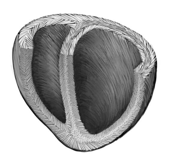
To build a human heart from the ground up, researchers need to replicate the unique structures that make up the heart. This includes recreating helical geometries, which create a twisting motion as the heart beats. It’s been long theorized that this twisting motion is critical for pumping blood at high volumes, but proving that has been difficult, in part because creating hearts with different geometries and alignments has been challenging.
Now, bioengineers from the Harvard John A. Paulson School of Engineering and Applied Sciences (SEAS) have developed the first biohybrid model of human ventricles with helically aligned beating cardiac cells, and have shown that muscle alignment does, in fact, dramatically increases how much blood the ventricle can pump with each contraction.
 This advancement was made possible using a new method of additive textile manufacturing, Focused Rotary Jet Spinning (FRJS), which enabled the high-throughput fabrication of helically aligned fibers with diameters ranging from several micrometers to hundreds of nanometers. Developed at SEAS by Kit Parker’s Disease Biophysics Group, FRJS fibers direct cell alignment, allowing for the formation of controlled tissue engineered structures.
The research is published in Science.
“This work is a major step forward for organ biofabrication and brings us closer to our ultimate goal of building a human heart for transplant,” said Parker, the Tarr Family Professor of Bioengineering and Applied Physics at SEAS and senior author of the paper.
Solving a 300-year-old mystery
This work has its roots in a centuries old mystery. In 1669, English physician Richard Lower — a man who counted John Locke among his colleagues and King Charles II among his patients — first noted the spiral-like arrangement of heart muscles in his seminal work Tractatus de Corde.


